All published articles of this journal are available on ScienceDirect.

Asbestos Bodies Burden in the Autopsy Lung Tissue from General Thai Population
Authors Info & Affiliations
Abstract
Background:
Chrysotile asbestos has been used in Thailand for over 30 years mainly in asbestos-cement wall and roof tiles. In non-exposed subject, asbestos fiber can contaminate in ambient indoor and outdoor environments.
Objective:
The aim of the present study is to evaluate the current prevalence and volume of AB load in general Thai population.
Methods:
Lung tissues were obtained from 200 autopsy cases. Asbestos Bodies (AB) were identified with light microscopy using the tissue digestion and membrane filtration method. Results are reported as AB/g wet lung tissue.
Results:
AB was identified in 97(48.5%) out of 200 cases. The AB level ranged from 0.19-14.4 AB/g wet lung. Most of the positive cases (99%) have less than 10 AB/g wet lung. Only one case exhibited a high value at 14.4 AB/g wet lung. Age, gender, occupation and hometown were found to have no effect on AB burden in autopsy lung tissue from this study.
Conclusion:
The prevalence of AB in autopsy lung tissue from general Thai population is 48.5% and the AB level ranges from 0-14.4 AB/g wet lung in consistent with non-occupational asbestos exposure level regarding several reference reports.
1. INTRODUCTION
Asbestos, a mineral silicate fiber, has been imported for industrial use in Thailand for over 30 years at a cost of 2-4 million USD yearly. Half of the imported asbestos is utilized by brake and clutch pads factories with the remaining primarily been used in the production of asbestos-cement wall and roof tiles. Initially, both chrysotile and amphibole asbestos fibers were imported. However, following numerous worldwide studies demonstrating an association between exposure to amphibole asbestos and respiratory diseases and mesothelioma, crocidolite and amosite were banned from Thailand in 1995 and 2001. This was followed by the prohibition of the import of actinolyte, anthophyllite and tremolite in 2009 [1]. Chrysotile asbestos is the only type of fiber that is currently allowed for use in industrial applications.
Generally, exposure to asbestos fibers can occur in occupational and environmental situations. In Thailand, occupational exposure typically occurs in workers during the manufacturing of asbestos-containing products. A study from the Occupational Safety and Health Bureau, Department of Labour Protection and Welfare indicated that asbestos fibers in the air from various areas of factories using asbestos are within the normal acceptable standard limit [1]. The study from Incharoen et al. [2] found that the asbestos body burden in bronchoalveolar lavage fluid from occupationally exposed workers was not significantly different from the general population.
In terms of non-occupational exposure, asbestos fiber can contaminate in ambient indoor and outdoor environments. Asbestos fiber can be found from weathering and erosion of asbestos-containing rocks and road surfaces, tears in brake and clutch pads, weathering of asbestos cement walls and roofing, especially during maintenance, repair and removal processes. Previously, studies from several industrial countries have demonstrated that the percentage of the Asbestos Body (AB) identified using the lung tissue digestion method from general autopsy cases varied from 16.4% to 100% [3-22], with an increased prevalence in the same population over time [4]. In Thailand, Sri-umpai et al. [5] identified AB in 33% of autopsy lungs since 1985.
The present study aims to evaluate the current prevalence and volume of AB load in autopsy lung representing the general Thai population. The effect of age, gender, occupation, and residential area to AB level will also be analyzed.
2. MATERIALS AND METHODS
2.1. Subjects
Samples from 200 autopsies performed at the Department of Pathology, Faculty of Medicine Ramathibodi Hospital, Mahidol University between September 2015 and September 2016 were included in the study. All experiments involving human subjects were approved by the Committee on Human Rights Related to Research Involving Human Subjects Faculty of Medicine Ramathibodi Hospital, Mahidol University, Thailand (Protocol number ID 07-58-41). Data from subjects including age, gender, occupation, hometown and cause of death were collected from the autopsy report.
2.2. Specimen Collection and AB Detection
The lung tissue was taken from both lower lungs and fixed in 10% neutral buffered formalin. AB was detected using the sodium hypochlorite digestion method from Smith and Naylor [23]. Briefly, the formalin-fixed lung tissue (approximately 5 g) was weighed and minced into 2-3 mm cubes. Then, the tissue was digested with 250 ml of sodium hypochlorite solution overnight or until no visible pieces of lung tissue. The supernatant was removed and 20 cc of chloroform and 50% ethanol were added to the sediment to removed pigments and lipid residues. The specimen was then centrifuged at 200 g for 10-15 minutes. The supernatant from each sample was removed until only 5 ml sediment remained. The sediment is suspended in 15 ml of 95% ethanol and filtered through 0.4-μm pore size, 25-mm-diameter Whatman Nuclepore® filter using Whatman Nuclepore® filtering apparatus (Sigma-Aldrich, MO, USA). The filter was transferred to a glass slide and left until dry. Chloroform was added dropwise until the filter was cleared. After evaporation of the chloroform, the coverslip was added with mounting media. The morphology of AB is a thin translucent core coated with golden brown beads, segmented or dumbbell shape [24]. The AB was counted from the whole slide under a light microscope, 200x and 400x magnification. AB burden was reported as AB/g wet lung.
2.3. Statistical Analysis
Comparison of mean AB/g wet lung between genders, occupational subtype and hometown was analyzed by independent T-test. The Pearson correlation coefficient was analyzed between age and AB/g wet lung. All statistical analyses were performed using SPSS version 18.0 (SPSS, Chicago, IL) and the statistical significance was set at p < 0.05.
3. RESULTS
From the 200 autopsy cases in this study, 136 were male and 64 were female with age ranging from 3 months to 88 years. From the autopsy report, data for occupation and residential area are available in 149 and 175 cases, respectively. The causes of death are present in Table 1. Occupation, hometown and level of AB for each group are described in Table 2.
| Cause of Death | Number of Cases (%) |
|---|---|
| Accident | 57 (28.5) |
| Myocardial infarction | 36 (18) |
| Infectious/sepsis/septic shock | 22 (11) |
| Others heart diseases | 21 (10.5) |
| Hanging | 13 (6.5) |
| Tumor-related | 11 (5.5) |
| Respiratory failure | 9 (4.5) |
| Pulmonary embolism | 5 (2.5) |
| Liver failure | 4 (2) |
| Homicide | 4 (2) |
| Hypovolemic shock | 4 (2) |
| Drowning | 3 (1.5) |
| Drug abuse | 2 (1) |
| Intracerebral hemorrhage | 2 (1) |
| Severe burn | 1 (0.5) |
| Epilepsy | 1 (0.5) |
| Malignant HT | 1 (0.5) |
| Pancreatitis | 1 (0.5) |
| Senility | 1 (0.5) |
| Unknown | 1 (0.5) |
| - | Number (%) | Number of AB+ Cases (%+) | AB/g Wet Lung |
|---|---|---|---|
| Occupation data (N=149)+ General employee2 Housewife1 Student and child1 Trader1 Government officer1 Private business1 Police2 Soldier2 Steward1 Salesman2 |
78 (38.5) 17 (8.5) 15 (7.5) 13 (6.5) 12 (6) 8 (4) 3 (1.5) 1 (0.5) 1 (0.5) 1 (0.5) |
37 (47.4) 10 (58.8) 3 (20) 5 (38.5) 5 (41.7) 7 (87.5) 1 (33.3) 1 (100) 1 (100) 1 (100) |
0.19-14.4 0.2-6.4 0.2-1.7 0.2-2.8 0.2-9.4 0.2-1 0.2 0.2 0.23 0.2 |
| Hometown (N=175)+ Bangkok3 Central4 Northeast4 South4 North4 East4 |
87 (49.7) 30 (17.1) 38 (21.7) 11 (6.3) 6 (3.4) 3 (1.7) |
42 (48.3) 15 (50.0) 18 (47.3) 7 (47.4) 3 (50.0) 2 (66.7) |
0.19-14.4 0.19-4.4 0.2-2.8 0.2-2.3 0.2-1 0.2 and 2.2 |
| - | Number (%) | Mean AB/g Wet Lung + SD | p-value |
|---|---|---|---|
| Gender (N=97) Male Female |
63 (64.9) 34 (35.1) |
1.29+ 1.9 1.05+ 1.9 |
0.74 |
| Occupational type* (N=72)+ Indoor Outdoor |
32 (44.4) 40 (55.6) |
1.17+ 1.9 1.25+ 2.4 |
0.88 |
| Hometown* (N=87)+ Bangkok Others provinces |
42 (48.3) 45 (51.7) |
1.55+ 2.7 0.86+ 0.9 |
0.12 |
AB was detected in 97 out of 200 subjects (48.5%). These were identified 46% and 53% of the male and female subject, respectively. Among the 97 positive cases; AB was identified ranging from 0.19-14.4 AB/g wet lung, 73 cases (75.3%) have <1 AB/g wet lung, 23 cases (23.7%) have 1-10 AB/g wet lung and only one case (1%) has >10 AB/g wet lung.
The youngest positive subject was a 1.5 years old infant with an AB concentration of 1.7 AB/g wet lung. No AB was found in subjects aged less than 1 year. The highest level of AB in the lung tissues was 14.4 AB/g wet lung from a 60-year-old male general employee who lived in Bangkok and died from hanging.
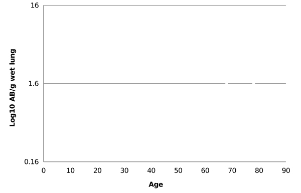
The mean and median AB/g wet lung tissue in 97 positive cases was 1.14 and 0.4, respectively. There was no correlation between age and AB/g wet lung, correlation coefficient (r) = 0.18 (Fig. 1). The mean AB/g wet lung with regard to gender, occupational subtype and hometown also showed no significant difference (Table 3).
4. DISCUSSION
The study of AB burden from autopsy lung tissue in general populations has been performed in many industrial countries throughout the world since 1960 until the current time [3-22]. The method for AB detection in lung tissue in early studies utilized a scraping technique [3, 6]. Subsequently, most analyses of AB burden shifted to use chemical digestion followed by AB collection through a membrane filter and detection based on observation under a light microscope [4, 5, 7-22]. The AB are counted and reported as AB/g dry lung, which is equal to 10 times of AB/g wet lung or AB/cm3. From the available data of these studies, the prevalence of AB identification ranges under 20% up to 50% in more recent studies and up to 80%-100% in the former studies [3-22]. The studies of the number of AB in lung tissue from various regions within countries or from different countries often show remarkable differences. Typically, AB was identified in less than 1000 AB/g of dry lung tissue or 100 AB/g of the wet lung in subjects who have no obvious history of asbestos exposure [25].
The long history of import and use of large amounts of asbestos in Thailand, 60,000 to 180,000 metric tons each year in the last 30 years [1], may be a risk factor for exposure. AB has been identified in 33% of subjects in a sampling of the general population lung tissue from Sri-umpai et al. 30 years ago [5]. In the current analysis, AB was identified in 97 out of 200 autopsy cases, which shows an increase in the prevalence of AB burden from 33% to 48.5%. In our opinion, most of the autopsy cases in this study lived in Bangkok, the capital city of Thailand, where the number of populations has significantly increased in the past 30 years. So, the more population means more air pollution, and poor air sanitation caused increase in the prevalence of AB comparing to the past. In term of AB level, both studies demonstrated that 99% of the positive cases have <10 AB/g wet lung (96 of 97 cases from the recent study and 108 of 109 cases from Sri-umpai et al.). The highest AB positive subject described by Sri-umpai et al. was 28.4 AB/g wet lung, while the current study was 14.4 AB/g wet lung. Even if the prevalence of AB in the autopsy lung has increased, the overall asbestos concentration in the general population in Thailand over the last 30 years tends to be similar, which is in contrast to some previous studies [7]. The most recent studies from Spain and Italy, which have completely banned asbestos for almost 20 years, show AB levels that exceed those identified in Thailand. Velasco-Garcia et al. from Spain identified AB in 86% of necropsy lung from the general population with <300 AB/g dry lung in all except one case with >1000 AB/g dry lung [8]. Casali et al. from Italy identified AB in 16.4% of necropsy subjects free from asbestos-related disease with concentrations ranging from 10-110 AB/g dry lung [9]. The recent study from Feder et al. demonstrates the persistent chrysotile fiber accumulation in the lung from asbestos-related disease subjects after exposure cessation for a long period of time [26]. Thus, AB appears to be present in autopsy lung from general populations whether that country has currently used or completely banned asbestos. This result may be due to insufficient clearance of fiber accumulate in the lung regardless of fiber type.
In terms of the age of each subject, we did not observe AB in subjects aged less than one year. The youngest positive subject was a 1.5 years old girl who died from congenital heart disease. This confirms that asbestos exposure can begin in the first year of life [27]. Although a study from Bhagavan and Koss demonstrated a higher AB concentration in older age group [10], several studies showed no increase in the number of AB with age [11, 12]. The result of our study also showed no significant correlation between age and level of AB/g wet lung. Occupation data also correlated with AB level especially outdoor occupation or “Blue Collar” is frequently associated with a higher level than an indoor occupation. The lower number has been reported in women compared to men, which is consistent with the greater chance for men to have occupational exposure to asbestos fiber [7, 10, 11, 16]. In this study, there is no gender predilection or associated type of occupation identified to relate with AB burden. The residential area also affects the AB burden in lung tissue, the level of AB appears to be higher in urban dwellers from some prior reports while the prevalence is similar between the urban and rural area from some reports [4, 7, 13]. In addition, no significant association between residential area and concentration of AB was observed in this study. However, the data was very limited in this study due to the lack of actual duration and the most recent data for occupational and residential information solely from autopsy reports.
CONCLUSION
AB was detected in 48.5% of individuals sampled from the Thai population with a concentration of 0.19-14.4 AB/g wet lung tissue. AB was identified in the lung of 1.5 year-old infant showing the potential for exposure at an early age. No significant correlation between age, gender or occupation and the asbestos burden was obtained from this study.
ETHICS APPROVAL AND CONSENT TO PARTICIPATE
This study was approved by the Committee on Human Rights Related to Research Involving Human Subjects Faculty of Medicine Ramathibodi Hospital, Mahidol University, Thailand (Protocol number 07-58-41).
HUMAN AND ANIMAL RIGHTS
No animals/humans were used for studies that are the basis of this research.
CONSENT FOR PUBLICATION
The informed consent was waived due to the leftover specimen included in the study.
AVAILABILITY OF DATA AND MATERIALS
The data sets used and/or analyzed during the current study are available from the corresponding author.
FUNDING
None.
CONFLICT OF INTEREST
The authors declare no conflict of interest, financial or otherwise.
ACKNOWLEDGEMENTS
The authors would like to thank Associate Professor Laran Jensen from Department of Biochemistry, Faculty of Science, Mahidol University, Bangkok, Thailand for manuscript writing support.

